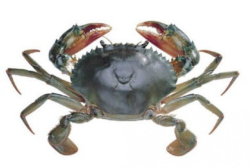
Mangrove crabs are one of the important valuable commodities found in mangrove and estuary areas. In Indonesia, consumers’ demand for mangrove crabs continues to increase due to a pattern of public consumption that shifts to the consumption of fisheries commodities. Moreover, the protein contents of mangrove crab include 44.85-50.58% protein, 10.52-13.08% fat, and energy 3.579-3.724 kcal / g (Karim, 2005). Mangrove crabs also contain omega-3 fatty acids and minerals that are good for the body.
Previous studies have found pathogen infestations from parasitic groups on mangrove crabs, including Zoothamnium, Epistylis and Octolasmis. The parasites were found attached to the gills and carapace of mangrove crabs.
One of the parasites that infest the mangrove crab and is macroscopic (visible to the eye) is Octolasmis. These parasites are found to infest gills or carapace in the form of sprouts with the head like a pear and have a tail (peduncle) that sticks in the body of mangrove crabs.
The emergence of ectoparasites can be an opportunity for other pathogens to multiply and one of them is derived from a group of fungi. This is evidenced by the presence of Lagenidium and aspergillus niger infections in mangrove crabs infested with Octolasmis ectoparasites.
Lagenidium sp. is a type of fungus that forms colonies like cotton with gray color. Lagenidium sp. has asexual reproduction by producing zoospores from hyphae that make vesicles.
Lagenidium sp. have long hyphae and round vesicles. Lagenidium sp. has coenocytic mycelium and hyphae that are strong, branched, with thin septa. The septa hypha is divided into several segments, which sometimes narrow to the septa. Segments sometimes branch off and separate from each other, called sub-thallus. Vesicles form in the final hypha, which contain a lot of sporangium produced by mycelium. Lagenidium sp. can be identified in SDA ( Sabouraud Dextrose Agar ) media macroscopically from the color and shape of the colony which is like cotton and gray.
Aspergillus niger can be identified in SDA ( Sabouraud Dextrose Agar ) media by the appearance of blackish-brown conidiophores. Microscopically, A. niger has the characteristics of transparent conidiophores and round and black sporangium.
The presence of bacterial infections in mangrove crabs as secondary infection can be caused by bacterial infections, parasitic infestations or unbalanced interaction of environmental components.
Author : Putri Desi Wulan Sari
More details of the article available on: