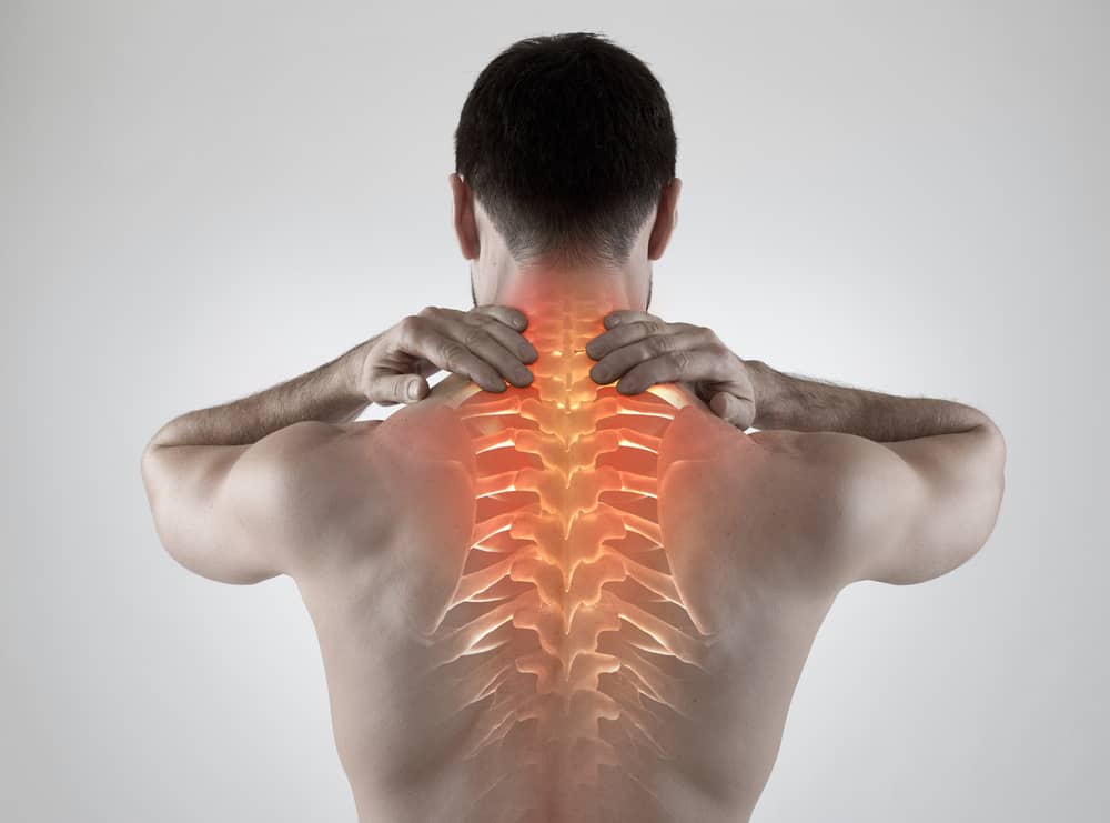
Schwannoma is a type of benign tumor on the cervical vertebrae that is rarely found. The incidence rate is only about 0.3-0.4 cases per 100,000 people. This tumor is located in the intradural region of spinal nerve and is a nerve sheath tumor originating from schwann cells. The existence of this tumor causes disruption to the body, from disruptive pain of the motor and sensory systems to even cause paralysis. This type of tumor mostly attacks people aged 40-50 years. Tumors that are more than 2.5 cm can be called “giant schwannomas”, where the difficulty of resection depends on the location and size of the tumor.
A case found at Dr. Soetomo was from a man, aged 54 years with a background in mechanical service. The patient first came to the clinic in August 2017 with difficulties in writing and coordination on the hands which caused disruption in doing his work. The patient had a history of tumor in the front neck four years ago and had removal surgery by a general surgical colleague, and a lymph node tumor was found. Complaints were felt since 6 months before coming to RSUD Dr. Soetomo and getting more severe over time. Neurological examination found a thick feeling below the C4 level bilaterally.
Comprehensive examination is carried out to find the cause of complaints in this patient, starting from a photo of the cervical vertebrae, to sophisticated MRI examination and angiography to determine the type of tumor in this patient. On plain photo examination, lytic lesions were found in C2 to C4 with bone damage in C3 and C4. CT scan obtained additional information in the form of a source of tumor arteries originating from the vertebral artery with the emphasis on the arteries causing stenosis until the diameter decreases, which is only 33% of the normal size of the vertebral artery. An MRI examination was performed and a 39.5 x 30.9 x 45.7 mm mass was obtained which indicated that this tumor was in Giant Schwannoma category that had invaded the intracanal region with scalloping destruction in neural foramina with expansion to the anterior and posterior.
After obtaining a fairly clear description of the disease and carried out discussions with various disciplines, it was decided to do surgery on this patient. The operation was carried out in two stages. The first surgery was performed from the back by removing the tumor and installing the implant. Tumor cells are taken using a CUSA® or aspirator ultrasonic cavitron, which is a special tool for removing tumor or cancer cells without damaging other surrounding tissue. Tumor cells were also taken a little to be examined in the anatomic pathology section. A fusion in the neck bone was done using instrumentation.
The second stage of the surgery was carried out from the front of the neck with the collaboration of head and neck surgeon and was performed three weeks later. Initially it was planned to cut the jawbone so that it could reach the C2 cervical vertebrae, but during the operation, it was not necessary to carry out such an action because the surgical instrument had already reached the tumor. The operation is carried out carefully not to damage the laryngeal nerve which is an important nerve for the vocal cords. After excision of the tumor, the pelvic bone graft is taken and a C2-C5 transplant with an anterior side plate. Postoperatively, the patient is placed in a cast from head to neck for 2 months to ensure stability of the operation.
Periodic evaluations continue to be carried out on patients. Within 3 months, the patient has regained his motor strength to 5/5 and can return to work as a normal mechanic. A case of upper cervical schwannoma tumors is quite rare, but do not rule out the possibility of good surgery and provide adequate satisfaction for patients. Department of Orthopedics and Traumatology Hospital Dr. Soetomo / Faculty of Medicine, Universitas Airlangga was able to handle this case well and with good planning and involved various multi-disciplines.
This case is only a small part of tumor and cancer cases handled by us. In the future, the handling of tumor cases will continue to evolve with times, and it is hoped that the introduction and early detection will further develop and the public will entrust the best treatment to Dr. Soetomo / FK Universitas Airlangga.
Author: Primadenny Ariesa Airlangga, dr., M.Si, Sp.OT(K)
Dep. Orthopedics and Traumatology, Faculty of Medicine, Universitas Airlangga Journal Title: Schwannoma of the upper cervical spine-a case report Authors: Primadenny Ariesa Airlangga, Bambang Prijambodo, Aries Rachmat Hidayat, Steesy Benedicta. Published in: Chinese Journal of Traumatology. 2019; 22 (6): 368-372 Link: https://www.sciencedirect.com/science/article/pii/S1008127519300653