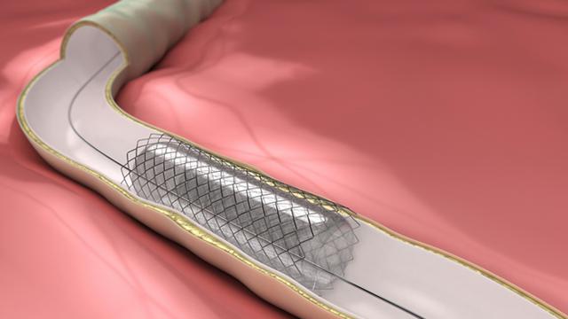
Heart disease is the highest silent killer in the world. In 2035, as much as 45% of the adult population in the United States will be affected by heart disease. Heart disease is closely related to atherosclerotic events, the narrowing of blood vessels due to plaque buildup in the walls of the heart. The most effective treatment for acute atherosclerosis is Coronary Artery Bypass Grafting (CABG) surgery, which is the replacement of narrowed arteries.
The vessels used in the CABG process are taken from other body parts such as the saphena magna vein and arteria thoracica interna. Unfortunately, the availability and compatibility of replacement vessels that can be used are quite limited and require surgical removal. Therefore, synthetic replacement vessels are needed as an alternative solution.
Synthetic vessels must meet the requirements to be well received by the human body. Some conditions, such as biocompatibility, hemocompatibility, biodegradable, non-toxic, non-clumping, and have excellent mechanical characteristics, need to be owned by synthetic vessels. Poly L-lactic acid (PLLA) polymer is one of the materials used to make synthetic vessels. PLLA is known as a biodegradable synthetic polymer, meaning that it can decompose automatically in the human body after a certain period.
The human body can accept PLLA because it contains lactic acid. However, PLLA does not support the growth of new cells around it. This cell growth factor needs to be added to PLLA so that it can be accepted and integrated with the surrounding cells. Besides, PLLA is hydrophobic, which is not binding to water. This property makes PLLA inhibited to merge with the surrounding cells. So, it is necessary to add individual components on the surface of PLLA to overcome this problem.
Chitosan is a biopolymer component that can be utilized in various fields. Chitosan was added to the PLLA surface to reduce the hydrophobic nature of PLLA. The structure of chitosan, which is similar to the surface structure of human cells makes chitosan accepted by the surrounding cells, even increasing the rate of cell growth. Chitosan is useful in cell regeneration and can be broken down and absorbed by the body naturally.
Collagen is a significant component of the extracellular matrix, which increases the interaction and elasticity of the surrounding cells. Collagen also has high toughness. Collagen, which has been known as one of the essential components in the cosmetics world, acts as a necessary component of the PLLA mixture. In a study conducted by Widiyanti et al, it was found that collagen and chitosan influence the heart ring characteristics.
The study was conducted to determine the effect of the addition of chitosan to the surface of the hollow fiber PLLA-shaped hollow (hollow tube). Five variations in the concentration used in the study with parameters such as hemocompatibility, the quality of interaction of the material with blood, and the cytotoxicity or the potential of the material to be toxic.
Hollow PLLA-collagen fiber is made by dissolving PLLA with 1% collagen using the spinneret apparatus method. The formed hollow fiber is coated with chitosan by immersing it in chitosan solution for 30 minutes. The hollow fiber is then dried using the freeze-drying method. 5 variations of the chitosan used were used, that is 0.05%; 0.075%; 0.1%; 0.125% and 0.15%. There is one hollow fiber that is not coated with chitosan as a comparison. Five hollow fiber samples were obtained with a smooth surface and had a thickness of 90.65 – 236.26μm.
After the hollow fiber is formed, a cytotoxicity test is performed on each sample to determine the level of the potential toxicity of material to human body cells. From the cytotoxicity test, the percentage of living cells in all samples exceeds 85%, with a minimum requirement of 50%. The sample with the lowest percentage was obtained from the sample with the highest chitosan concentration. Cytotoxicity test results indicate that all hollow fiber samples used are non-toxic, and the surrounding cells can live well.
Hemolysis test is performed to determine the degree of material haemocompatibility. From the hemolysis test, the percentage of hemolysis in all samples is less than 3.3%, with a maximum requirement of 5%. The sample with the highest percentage is the sample with the lowest concentration of chitosan. The percentage of hemolysis decreases with increasing chitosan concentration. Hemolysis test results indicate that all hollow fiber samples used can interact well with the blood.
Based on the research, it can be concluded that the addition of chitosan concentration has a positive effect on hollow fiber PLLA-collagen by decreasing the percentage of hemolysis and increasing the hemocompatibility of a material. The addition of chitosan layer makes the PLLA-collagen hollow fiber surface smoother so that it can reduce the friction between the surface of the hollow fiber and the flowing blood. There is potential for further research to determine the effect of the addition of chitosan to the process of endothelial cell formation in artificial blood vessels.
Author: Prihartini Widiyanti
Link:
http://jurnalnasional.ump.ac.id/index.php/medisains/article/view/5259/0