As one of the deadliest infectious diseases in the world, tuberculosis made Indonesia has the second highest number of TB cases after India in 2018. TB predominantly affects the lungs and is categorized as pulmonary TB. However, it could also occur in other sites of the body, such as the joints, meninges, bones, and lymph nodes, including oral cavity. Although its incidence is approximately 0.05%–5% of total TB cases, dental practitioners should pay attention to it.
To date, problems often reported by dental practitioners are oral cavity TB with varied clinical presentations, such as oral ulcer, nodules, gum inflammation and lesions; these lesions could be both painful or painless. All of these factors could result in misdiagnosis. Dentists could play a vital role in the early detection of oral TB lesions to ensure its prompt management.
This study aims to assess and identify the oral lesions, both tubercular and nontubercular, of patients with pulmonary TB in outpatient clinic of Dr. Soetomo General Hospital Surabaya, East Java, Indonesia from July 2017 to January 2018. From 30 patients of pulmonary tuberculosis, 24 patients have oral cavity lesions and most of them were in their tongues.
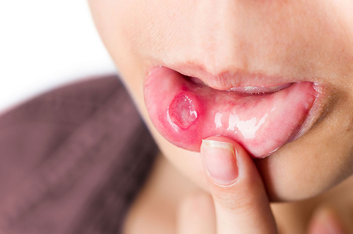
Among 37 lesions found from the subjects, only one lesion related with tuberculosis (tubercular lesion), from the lesion characteristics; it was a single approximate size ± 2 cm, irregular border, yellowish, and painless ulcer on the ventral part of the tongue.
Coated tongue, a condition of white or brownish yellow layers on the surface of the tongue, is the lesion with the most frequency. The second most cases is crenated tongue, which is a hollow form like a dental mold on the edge of the tongue, whereas the third most cases are fissured tongue, cracks / grooves on the surface of the tongue as if the tongue looks broken.
In addition to the aforementioned cases, two patients were found to have a fungal infection in the oral cavity, which was marked by white patches of fake membranes and could be scraped off the tongue and reddish spots on the palate. This fungal infection is usually called Oral Candidiasis, which is often associated with systemic diseases, especially immunocompromised conditions, such as HIV / AIDS. The reference states that the incidence of Candida infection in tuberculosis patients ranges between 15 – 32%.
It shows that the majority of pulmonary tuberculosis patients have lesions in the oral cavity, however, oral cavity lesions associated with tuberculosis are rarely found. Coated tongue lesions are common, although in principle it is a normal variation of the oral cavity, often associated with a systemic disease condition, as these conditions result in changes in the oral cavity. Because some lesions may appear without symptoms, routine dental examinations by the dentist certainly play an important role in detecting early oral cavity abnormalities for better treatment.
Author: Reiska Kumala Bakti
Details of this study available at:
http://www.jidmr.com/journal/wp-content/uploads/2019/12/54.19153-ED-2-OK_fl.pdf