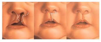
The goal of cleft lip treatment is to restore the symmetrical and natural shape of the lips and nose without any disruption in function. Treatment is carried out with an approach to any anatomic and pathological abnormalities. However, deformity and asymmetry are still found after the cleft lip surgery and it requires secondary correction.
Postoperative assessment after a cleft lip surgery is generally done using two-dimensional (2D) and three-dimensional (3D) photographs. In the case of unilateral cleft lip, it is difficult to determine the center line of the lips, which is important for evaluating the level of facial symmetry.
In the field of medical research, software to analyze and simulate the morphology of the body before and after surgery was developed using a non-contact 3D type measuring device. 3D analysis of lip and nose shapes before and after surgery can show changes in facial structure, and can predict the outcome of surgery, regardless of the operator’s skill and experience.
The upper lip skin surface indicates the success of the operation, but it is very difficult to assess the surface asymmetry of young people’s faces, especially those that are not cooperative during data acquisition. The development of 3D stereophotogrammetry allows us to obtain precise 3D data on the surface of young people after the primary cleft lip surgery.
In this study, we propose an image processing method for visual and quantitative assessment of facial surfaces digitally using shadow and zebra images, which are constructed from moire images reconstructed based on 3D facial data. Furthermore, we compared the upper lip shape asymmetry in cleft lip patients and normal children to assess the muscle reconstruction techniques that we applied when closing the primary lip gap.
Twenty-two complete unilateral cleft lip patients from October 2009 to January 2013 were involved in this study. All patients had undergone primary closure surgery with the Cronin method (Cronin, 1966) at an average age of 5.1 + 1.6 months. 3D facial data from patients were taken using stereophotogrammetry, Vectra H1, mean age 64.0 + 11.5 months. 3D control data values were obtained from 23 healthy children of the same age and gender distribution.
Image shadow and zebra constructed from images moire , reconstructed from 3D face data using stereophotogrammetry. The image is then cropped and reversed with a central axis of the face. The difference in center of gravity and density between the cleft side of the lips and the healthy side in the two attractive areas, namely the face and the lip area are calculated and compared with the control values in healthy children.
In the group of cleft lip patients, the mean shadow and zebra image discrepancies were 1.76 +/- 0.70 and 2.63 +/- 1.72 pixels, respectively, in the face area and 1.31 + / – 0.36 and 3.83 +/- 2.08 pixels, respectively, in the lip area. There was a significant difference in the mean center of gravity and density in the zebra image in the lip area between the lip gap and the control group.
In conclusion, our image analysis of digital facial surface asymmetry in patients with complete unilateral cleft lip can provide visual and quantitative information, and it can contribute to the improvement of muscle reconstruction in cleft lip surgery. (*)
Author: Muhammad Subhan Amir
Details of research available at: