Bone damage caused by accidents, tumors or joint reconstruction often occurs, and material that can replace the injured bone is needed. There are several types of synthetic material implanted in the damaged part of the bone, but they still cause encapsulation by fibrous tissue and fail to unify with the original bone. Osteoporosis is one type of bone damage characterized by a decrease in bone strength caused by reducing bone density. Osteoporosis is also one of the main risk factors for bone fractures. World Health Organization(WHO) in 2013 reported that around 200 million people in the world have osteoporosis, and 50% of them suffer bone fractures, especially in the upper legs or femur. Bone damage is generally caused by external factors, but osteoporosis is caused by internal factors, such as the declining ability of bones to perform bone remodelling due to unbalanced work between osteoblasts, which form bone cells, and osteoclasts, which works for bone remodeling. The work of the osteoclasts exceeds the work of osteoblasts, so the process of bone remodeling is faster than the process of bone cell formation. The result of this unbalanced work is a declining bone density.
The method that can be used to overcome this situation is generally by taking drugs that work by inhibiting osteoclast work in the process of bone remodeling. It can also be done by increasing bone density using substitute material, as mentioned earlier. One type of material that can replace the role of bone and is widely used as bone filler is hydroxyapatite. This material is bioactive and has been explored to treat bone damage due to osteoporosis. Unfortunately, this material is quite brittle and requires other materials as a mixture to compensate for these weaknesses. One of the materials that can be used is gelatin. The addition of gelatin aims to improve adhesion, migration and mineralization of osteoblasts.
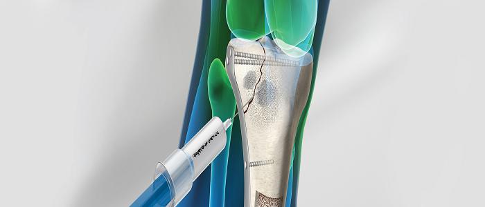
The use of hydroxyapatite and gelatin as bone filler is not very effective because of its dense shape so bone filling material is developed in the form of injectable or commonly called the Injectable Bone Substitute (IBS). By using IBS, the bone filling material can adjust to irregular shaped bone damage due to osteoporosis.
IBS itself is generally divided into two types: IBS which is ready to be used in the form of suspension and IBS which is composed of ionic hydraulic cement that can harden in vivo after injection. Previous studies have reported that HPMC is often used as a forming suspension agent in IBS. Also, this IBS function can function as a drug delivery agent, in this case, the medicine for osteoporosis, alendronate. Alendronate has high electron affinity for calcium ions, which can increase interaction with bone calcium and inhibit osteoclasts in the process of bone remodeling.
This study was able to synthesize and characterize the hydroxyapatite-gelatin composite suspension with the addition of alendronate as IBS. The in vitro characterization carried out on the suspension included the Fourier Transform Infrared (FTIR), viscosity, resuspension, acidity (pH), cytotoxicity, and setting time test. The suspension is made by stirring hydroxyapatite and gelatin 5% (w / v) with a ratio of 40:60, 45:55, 50:50 and 55:45 and alendronate as much as 10% of the hydroxyapatite used. The mixture of hydroxyapatite, gelatin and alendronate is mixed with HPMC 2% (w / v). The FTIR test results showed the formation of a bond between hydroxyapatite and gelatin as indicated by the shift of the carboxyl group absorption area from gelatin in the area of 1332.72 cm-1 becomes 1559-1543 cm -1 , the bond between the carboxyl group (COO – ) of gelatin with Ca 2+ from hydroxyapatite. The best composition variation of suspension as IBS is hydroxyapatite: gelatin = 45:55 with viscosity value (38.7 ± 0.53) dPa.s, acidity (pH) reaches 7 and cell viability percentage more than 50%. The resuspension test results showed that the suspension formed a suspension again after deposition and did not change the pH of the Simulated Body Fluid(SBF) solution. The setting time test results show that the suspension is setting with the condition that it must be on the appropriate substrate. Scaffold Hydroxyapatite-collagen was used as a substrate, and the results showed an increase in pore size with a range ranging from 17,271 µm to 21,316 µm starting from 8,052 µm to 11,631 µm from the results of Scanning Electron Microscope (SEM) test.
The conclusion of this study shows that IBS based on hydroxyapatite and gelatin with a ratio of 45:55 and the addition of alendronate can support its application as bone filler that delivers drugs for osteoporosis cases.
Author: Alfian Pramudita Putra, S.T., M.Sc.
Complete information about this research can be seen in our publication in the Journal of International Dental and Medical Research (Q3) at the following link:
http://www.jidmr.com/journal/wp-content/uploads/2019/07/76_M18_706_Alfian_Pramudita_Putra_Layout.pdf
A. P. Putra, D. Hikmawati, A. S. Budiatin, “Injectable Bone Substitute of Hydroxyapatite-Gelatin Composite with Alendronate for Bone Defect Due to Osteoporosis”, J. Intl. Dent. Med. Res. 2: pp. 813-816. (2019).